rAlbumin Human
If you find any inaccurate information, please let us know by providing your feedback here

Tóm tắt nội dung
This article is compiled based on the United States Pharmacopeia (USP) – 2025 Edition
Issued and maintained by the United States Pharmacopeial Convention (USP)
DOWNLOAD PDF HERE
1 DEFINITION
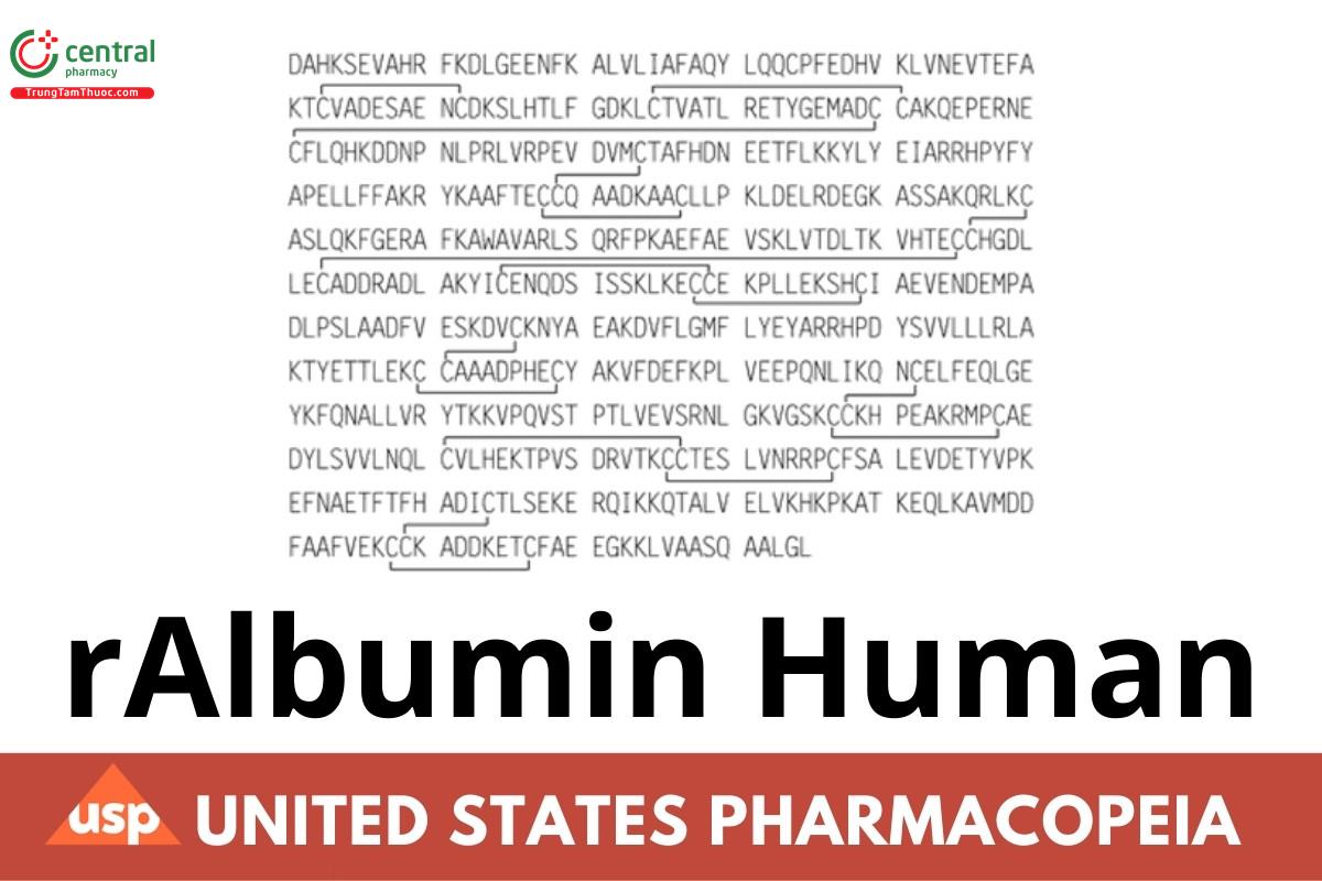
Recombinant Albumin Human (rAlbumin Human or rHA) is produced by recombinant DNA expression in Saccharomyces cerevisiae. Structural equivalence (primary, secondary, and tertiary) between rHA and human serum albumin (HSA) has been demonstrated. It consists of three domains composed of 585 amino acids containing a single tryptophan (Trp ), one free thiol (Cys ), and 17 disulfide bridges. It is presented
as a sterile and nonpyrogenic aqueous liquid consisting of a 10% (w/v) or 20% (w/v) solution in Water for Injection. No human- or animal- derived raw material is involved in its manufacture. It contains NLT 95% and NMT 105% of the labeled amount and NLT 99% of its total protein is albumin. It contains no added antimicrobial agents, but it may contain appropriate stabilizing agents. The presence of process- related impurities, host cell DNA, and host cell proteins is process specific; suitable limits should be determined by appropriately validated methods. However, the limit for host cell proteins should be NMT 0.15 µg/g.
2 IDENTIFICATION
2.1 A. PEPTIDE MAPPING
[NOTE—See Biotechnology-Derived Articles—Peptide Mapping 〈1055〉 for guidance.]
Tris buffer: 0.1 M tris(hydroxymethyl)aminomethane. Adjust with hydrochloric acid to a pH of 8.0. Dilute Tris buffer: Tris buffer and water (50:50)
Solution A: Trifluoroacetic acid and water (1:1000)
Solution B: To 350 mL of acetonitrile add 150 mL of water and 425 µL of trifluoroacetic acid. Dithiothreitol solution: 0.1 M dithiothreitol
Iodoacetamide solution: 0.1 M iodoacetamide in Tris buffer
Trypsin solution: 1 mg/mL of trypsin in 10 mM hydrochloric acid EDTA solution: 0.1 M ethylenediaminetetraacetic acid (EDTA) in water
Diluent: To 5.76 g of guanidine hydrochloride add 5 mL of Dilute Tris buffer and 200 µL of EDTA solution. Dilute with Dilute Tris buffer to a final
volume of 10 mL.
Standard solution: Add 20 µL of USP rAlbumin Human RS to 80 µL of Diluent. Add 5 µL of Dithiothreitol solution, and incubate at 37° for 75 min. Add 10 µL of Iodoacetamide solution, and incubate for an additional 75 min at 37° in the dark. Add 100 µL of Dilute Tris buffer, 400 µL of water, and 10 µL of Trypsin solution, and incubate at 37° with shaking for 24 h. Centrifuge, and dilute a portion of the supernatant in Solution A (50:50).
Sample solution: 50 mg/mL of rAlbumin Human in water. To 20 µL of this solution add 80 µL of Diluent. Add 5 µL of Dithiothreitol solution, and incubate at 37° for 75 min. Add 10 µL of Iodoacetamide solution, and incubate for an additional 75 min at 37° in the dark. Add 100 µL of Dilute Tris buffer, 400 µL of water, and 10 µL of Trypsin solution, and incubate at 37° with shaking for 24 h. Pulse centrifuge, and dilute a portion of the supernatant in Solution A (50:50).
Mobile phase: See Table 1.
Table 1
Chromatographic system
(See Chromatography 〈621〉, System Suitability.) Mode: LC
Detector: UV 214 nm
Column: 4.6-mm × 25-cm; 5-µm packing L1 Column temperature: 35°
Flow rate: See Table 1.
Injection volume: 100 µL Analysis
Samples: Standard solution and Sample solution
Acceptance criteria: The peptide map chromatographic profiles of the Sample solution are similar to those of the Standard solution.
2.2 B. ELECTROSPRAY MASS SPECTROMETRY
Solution A: Trifluoroacetic acid and water (1:1000)
Solution B: To 140 mL of acetonitrile add 60 mL of water and 180 µL of trifluoroacetic acid. Solution C: Acetonitrile and water (50:50)
Solution D: To 5 mL of Solution C add 10 µL of formic acid.
System suitability solution: Dissolve 2 mg of horse heart myoglobin in 589 µL of Water for Injection. Dilute 25 µL of this solution with 475 µL of Solution D.
Sample solution: 10 mg/mL of rAlbumin Human in water
Mobile phase: See Table 2.
Table 2
Chromatographic system
(See Chromatography 〈621〉, System Suitability.) Mode: LC
Detector: UV 280 nm
Column: 2.1-mm × 3-cm; desalting cartridge, equilibrated with Solution C1
Flow rate: 0.2 mL/min
Injection volume: 20 µL of the Sample solution
Analysis: Desalt the Sample solution, and collect the eluate. Ensure that a single protein peak elutes.
Spectrometric system
(See Mass Spectrometry 〈736〉.) Mode: LC/MS (using infusion pump)
[NOTE—The infusion system flow rate can be adjusted as needed. To assist in nebulization, the infusion system can contain a sheathing gas fluid.]
Mobile phase: Solution C
Detector: Electrospray in the positive ion mode Injection volume: 50 µL of desalted Sample solution
System suitability
Sample: System suitability solution Suitability requirements
Peak position: A single peak in the 16,949–16,953 Da range is found.
Analysis: Obtain and transform the spectrogram for the desalted Sample solution. Acceptance criteria: The mass is within 20 Da of the theoretical mass.
3 ASSAY
Change to read:
3.1 ALBUMIN CONTENT
Stock sample buffer:2 Mix 4 mL of 0.5 M Tris hydrochloride pH 8.6, 0.5 mL of 0.1% bromophenol blue, 2.0 mL of Glycerol, and dilute with water to 1000 mL.
Diluted sample buffer: Stock sample buffer and water (1:1)
Native stock running buffer:3 29 mg/mL of Tris base and 144 mg/mL of glycine (ERR 1-Oct-2024)
Running buffer: Native stock running buffer and water (1:9)
Gel-staining solution: A suitable Coomassie G-250-based solution4 Native PAGE gel: Prepare a 14% Tris-Glycine gel.5
Sample solution: 4 mg/mL of rAlbumin Human in water. Dilute this solution with Stock sample buffer to 2 mg/mL.
Calibration solutions: Dilute the Sample solution quantitatively, and stepwise if necessary, with Diluted sample buffer to 100, 20, 15, 10, 5, 2, and 1 µg/mL of rAlbumin Human.
Electrophoretic system
Run buffer: Running buffer Voltage: 125 V
Amperage: 35 mA
Wattage: 5.0 W
Run time: Approximately 2 h Loading volume: 10 µL
Analysis
Samples: Sample solution and Calibration solutions Gel loading scheme
Lane 1: 1 µg/mL Calibration solution
Lane 2: 2 µg/mL Calibration solution
Lane 3: 5 µg/mL Calibration solution
Lane 4: 10 µg/mL Calibration solution
Lane 5: 15 µg/mL Calibration solution
Lane 6: 20 µg/mL Calibration solution Lane 7: Diluted sample buffer
Lane 8: Sample solution Lane 9: Sample solution
Lane 10: Diluted sample buffer
Gel staining: Place the gel in 100 mL of water, and shake gently with circumgyration for about 30 min. Pour approximately 50 mL of Gel- staining solution into a staining container. Place the gel into the staining container, and allow the stain to completely cover the gel. Place the staining container on an orbital shaker, and stain the gel for 120 min with gentle shaking.
Destaining: Drain the Gel-staining solution, and add 100 mL of water to the container to cover the gel. Place the container on an orbital shaker, and shake at low speed for about 60 min. Change the water, and repeat for a total of two washes.
Gel scan procedure: Set up a gel scanner according to the manufacturer's instructions. Place the gel in the detector, and obtain a single image of all 10 lanes of the gel.
Data analysis: Perform image analysis of Lanes 1–6 to generate a linear calibration curve. Determine the linear regression equation of the standards by the least-squares method, with standard concentrations, in ng, as the dependent variable (x), and the sample band intensity (optical density) as the independent variable (y). Record the linear regression equation and the correlation coefficient, r. A suitable system is one that yields a line having an r2 of NLT 0.990.
Examine Lanes 8 and 9 (the Sample solution lanes) for the presence of bands below the main albumin band. If bands are present below
the main albumin band in either or both lanes, quantify the relative amount, in ng, of protein present in each band against the calibration curve. Convert the quantified value to a contaminant level in percentage by dividing the quantified value by a factor of 200.
Calculate the purity of the Sample solution:
Result = 100 − Ci
Ci = mean of the percentages of contaminant levels found in Lanes 8 and 9 (all the bands other than the albumin band), disregarding any band due to the Diluted sample buffer
Acceptance criteria: Sample solution purity is NLT 99.0%. [NOTE—The main albumin band is not quantitated. See the test for Total Protein.]
3.2 TOTAL PROTEIN
Sodium chloride solution: 0.15 M sodium chloride in water
Copper sulfate solution: 60 mg/mL of copper sulfate pentahydrate and 600 mg/mL of potassium sulfate6 in sulfuric acid low in nitrogen Sample solution: Dilute 0.5 g of rAlbumin Human with 2.5 mL of Sodium chloride solution (equivalent to about 3.3 mg/mL of total protein). Blank: 33.3 mg/mL of glycine in Sodium chloride solution
Analysis: To 3.0 mL of the Sample solution and the Blank, in suitable distillation tubes, add 5 mL of Copper sulfate solution. Incubate at 420°
for a minimum of 2 h, or until the residues appear white. When the solutions are cool, transfer the residues quantitatively with a minimum quantity of water to a micro-Kjeldahl flask, and determine the residues, using Nitrogen Determination 〈461〉, Method II. Multiply the result, corrected for the Blank and for the specific gravity of the Sample solution, by 6.25 to calculate the quantity of protein.
Acceptance criteria: 95%–105% of the quantity of protein stated on the label
4 OTHER COMPONENTS
4.1 SODIUM CONTENT
Diluent: 1.0 mg/mL of cesium chloride in water
Standard solutions: Prepare 0.5, 1.00, 1.50, and 2.00 mg/mL of sodium chloride in Diluent. Sample solution: 80 µg/mL of rAlbumin Human in Diluent
Apparatus
Mode: Atomic absorption Emission wavelength: 589 nm
Analysis: [NOTE—Use peak area measurements for quantitation.] Samples: Diluent (as blank), Standard solutions, and Sample solution
Introduce a blank solution (Diluent) into the atomic generator, and adjust the instrument reading to zero. Determinations are made by comparison with the Standard solutions of known concentration. If the Sample solution emission exceeds that of the Standard solutions with the highest concentration, dilute the Sample solution with Diluent. Introduce the most concentrated Standard solution into the instrument, and adjust the sensitivity to obtain a suitable reading. Introduce the Sample solution and Standard solutions into the instrument at least three times, and record the steady reading. Rinse the apparatus with blank solution each time, and ascertain that the reading returns to its initial blank value. Plot the mean of the readings obtained for the Standard solutions against their respective sodium concentrations. From the standard curve, calculate the sodium concentration content in the Sample solution, and adjust for the specific gravity of the rAlbumin Human (see Total Protein).
Acceptance criteria: 120–160 mM sodium IMPURITIES
Change to read:
4.2 LIMIT OF HIGH MOLECULAR WEIGHT PROTEINS
Solution A: 200 mg/mL of sodium azide
Buffer: Dissolve 54.2 g of dibasic sodium phosphate dihydrate, 30.0 g of monobasic sodium phosphate dihydrate, and 284.0 g of anhydrous sodium sulfate in 1600 mL of water. Add 50 mL of Solution A, and dilute with water to 2000 mL.
Mobile phase: Buffer and water (10:90)
Sample solution: 40 mg/mL of rAlbumin Human Chromatographic system
(See Chromatography 〈621〉, System Suitability.) Mode: LC
Detector: UV 280 nm
Column: 7.8-mm × 30-cm; 5-µm packing L59 Flow rate: 1.0 mL/min
Injection volume: 50 µL. [NOTE—The peak due to high molecular weight impurities, such as the polymer of albumin, appears in the void volume of the chromatogram.]
Analysis
Sample: Sample solution
Calculate the percentage of albumin polymer in the sample:
Result = (rU/rT) × 100
rU = peak response of albumin polymer
rT = sum of all rAlbumin Human related peak responses
Acceptance criteria: NMT 1.0% (ERR 1-Oct-2024)
5 SPECIFIC TESTS
PH 〈791〉
Sample solution: 1% (w/v) protein solution diluted with 0.9% (w/v) sodium chloride Acceptance criteria: 6.4–7.4
STERILITY TESTS 〈71〉: Meets the requirements
BACTERIAL ENDOTOXINS TEST 〈85〉: NMT 0.5 USP Endotoxin Unit/mL of rAlbumin Human
6 ADDITIONAL REQUIREMENTS
PACKAGING AND STORAGE: Preserve in tight glass containers, and store at 2°–8°. Do not freeze.
LABELING: Label to indicate that the material is of recombinant DNA origin.
USP REFERENCE STANDARDS 〈11〉
USP rAlbumin Human RS
1 A suitable reverse-phase desalting column is available from Perkin Elmer (No. 0711-0056).
2 Available from Invitrogen (No. LC2673).
3 Available from Invitrogen (No. LC2672).
4 A suitable Coomassie stain is available from Pierce (No. 24890 or No. 24592).
5 Alternatively, a precast 14% Tris-Glycine gel is available from Invitrogen (No. EC6485).
6 Copper sulfate pentahydrate and potassium sulfate tablets (each tablet with 1.5 g of K SO + 0.15 g of CuSO · 5H O) are available from Foss (No. 15270054).
