RADIOACTIVITY-THEORY AND PRACTICE
If you find any inaccurate information, please let us know by providing your feedback here


Tóm tắt nội dung
- INTRODUCTION
- TYPES OF DECAY
- GENERAL CONSIDERATIONS
- Radioactivity
- Fundamental Decay Law
- Counting Efficiency
- Background
- Statistics of Counting
- Minimum Detectable Activity
- Limit of Quantification
- Counting Losses
- Linearity and Range
- Calibration Standards
- Production of Radionuclides
- Carrier
- Radiochemical Identity and Purity
- Radionuclidic Identity and Purity
- Chemical Purity
- Labeling
- Naming Conventions for Isotopes
- INSTRUMENTATION FOR DETECTION AND MEASUREMENT OF RADIOACTIVE EMISSIONS AND APPLICATIONS
- GLOSSARY
- REFERENCES
This article is compiled based on the United States Pharmacopeia (USP) – 2025 Edition
Issued and maintained by the United States Pharmacopeial Convention (USP)
1 INTRODUCTION
Radioactive drugs and devices require specialized techniques in their production, testing, handling, dispensing, and administration to ensure optimal effectiveness and maintain safety for workers, patients, and the public. All operations involving these articles should be carried out by or under the supervision of personnel who have been appropriately trained in the handling of radioactive materials.
The facilities for the production, storage, and use of radioactive drugs and devices are generally subject to licensing by the U.S. Nuclear Regulatory Commission, an appropriate state agency, or similar governmental agencies outside of the United States. Most radioactive drugs and devices, although not identified as hazardous drugs, are classified as hazardous materials and are therefore subject to other regulations relating to transportation, environmental release, and workplace safety.
The purpose of this chapter is to provide information regarding radioactivity-including definitions, types of decay, and general considerations relating to radioactive decay, counting, radionuclide production, purity, and labeling-as well as instrumentation for detection and measurement of radioactive emissions.
Specific information on standards for radionuclide identification and assay, including instrument qualification, performance checks, identification of radionuclides and radionuclidic impurities, and assay of radionuclides are provided in Radioactivity 〈821〉.
2 TYPES OF DECAY
“Radioactive decay” is the process by which an unstable nuclide transitions to a lower energy configuration. Depending on the particular starting radionuclide, the result of the transition may be either a stable nuclide or a different radionuclide. Typically, these transitions are accompanied by the emission of radiation from the nucleus, which is broadly classified as either particulate or nonparticulate. Some radionuclides emit multiple types of radiation in this process, whereas others emit only a single type. The main types of particulate radiation commonly seen in nuclear medicine are alpha, beta, and positron. Nonparticulate types of radiation include gamma rays and X-rays. Nuclear medicine imaging is accomplished through detection and localization of nonparticulate radiation, whereas therapeutic effects arise from the energy deposited in the target organ by particulate radiation.
2.1 Alpha Decay
“Alpha decay” is radioactive decay with the emission of alpha particles, or helium nuclei, and is generally limited to elements with an atomic number >83. In some cases, beta particles and gamma rays may also be emitted during alpha decay. An example of a radionuclide that decays by alpha decay is:
Radium-226 → Radon-222
2.2 Beta Decay
“Beta decay” is radioactive decay with the emission of an electron. This type of decay typically occurs in neutron-excessive radionuclides wherein a neutron is transformed into a proton. In some cases, the emission of a positively charged electron, or positron, may occur. This type of decay typically occurs in neutron-deficient radionuclides with a lower atomic number wherein a proton is transformed into a neutron. In some cases, gamma rays may also be emitted during beta decay. An example of beta decay through emission of an electron is:
Iodine-131 → Xenon-131
An example of beta decay through emission of a positron (β⁺) is:
Fluorine-18 → Oxygen-18
Because a positron is an anti-electron, when it interacts with an electron, the two particles annihilate, and their combined mass is transformed into energy in the form of two 511 kiloelectron volt (keV) gamma rays. These gamma rays are produced simultaneously and travel away from the point of interaction in nearly opposite directions. These two characteristics form the basis for positron emission tomography (PET) imaging techniques.
2.3 Electron Capture Decay
“Electron capture” is radioactive decay that involves nuclear capture of an inner orbital electron, nuclear transformation of a proton into a neutron, and emission of one or more gamma rays. Electron capture generally occurs in higher atomic number radionuclides that are neutron deficient. An example of a radionuclide that decays by electron capture decay is:
Iodine-123 → Tellurium-123
2.4 Isomeric Transition
An “isomeric transition” is radioactive decay that involves a transition between nuclear isomers with the emission of one or more gamma rays. In contrast to other types of decay, the number of protons and neutrons remains the same in an isomeric transition. Isomeric transition generally occurs in radionuclides that are metastable. An example of a radionuclide that decays by isomeric transition is:
Technetium-99m → Technetium-99
3 GENERAL CONSIDERATIONS
3.1 Radioactivity
Radioactive decay is a first-order process (i.e., a fraction of atoms decay per unit time). The rate of decay for each radionuclide is a unique and constant value, which gives rise to the term “decay constant”.
Each radionuclide's rate of decay is a unique and constant value (its decay constant) and can be described by the following equation:
A = λN
A = amount of radioactivity in a source at a given time
λ = rate of decay of the radionuclide
N = number of radioactive atoms
The traditional unit for radioactivity is the curie (Ci), which is equal to 3.7 × 10¹⁰ atoms undergoing radioactive decay, or disintegrations per second (dps). Commonly used prefixes associated with the Ci include the millicurie (mCi) and the microcurie (µCi). The SI unit for radioactivity is the becquerel (Bq), which is equal to 1 dps. Commonly, prefixes associated with the Bq include the megabecquerel (MBq) and the gigabecquerel (GBq). Hence, 1 Ci = 37 GBq.
3.2 Fundamental Decay Law
The decay of a radioactive source is described by the equation:
Nₜ = N₀e⁻λt
Nₜ = number of radioactive atoms remaining at elapsed time t
t = time elapsed (time unit, such as seconds, minutes, or hours)
N₀ = number of radioactive atoms when t = 0
λ = decay constant of the specific radionuclide
The above equation can be rewritten in terms of radioactivity:
Aₜ = A₀e⁻λt
Aₜ = amount of radioactivity at elapsed time (t)
t = time elapsed (time unit, such as seconds, minutes, or hours)
A₀ = amount of radioactivity when t = 0
λ = decay constant of the specific radionuclide
“Decay tables” that provide radionuclide-specific decay factors (i.e., fraction remaining) calculated from A₀e⁻ˡᵗ at various elapsed times (t) are commonly available.
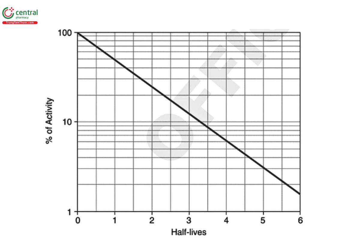
The half-life is defined as the time interval required for a quantity of radioactivity to decay to one-half of its initial value and is related to the decay constant λ by the equation:
T₁⁄₂ = 0.69315 / λ
T₁⁄₂ = half-life of the radionuclide
λ = decay constant of the specific radionuclide
The activity of a pure radioactive substance as a function of time can be obtained from the exponential equation, from decay tables, or by graphical means based on the half-life (Figure 1).
3.3 Counting Efficiency
The validity of any radionuclide measurement is dependent upon the reproducibility of the relationship between the source, the detector, and its surroundings. Appropriate allowances should be made for source configuration. Measurements of radioactivity require calculation of a calibration factor, or efficiency, and are dependent on the type of detector, the container in which the radioactivity is placed, and the source–detector geometry. The basic efficiency equation for detectors designed to count discrete decay events over a period of time is given as:
disintegrations/s = (counts/s) / ε
ε = efficiency or calibration factor
Because 1 dps is defined as 1 Bq, the above equation gives radioactivity in units of Bq. Through the application of the appropriate unit conversion, Bq can be transformed into µCi or any other unit of radioactivity. Detectors should be calibrated with a source of known radioactivity and in a fixed geometry.
3.4 Background
Cosmic rays, radioactivity present in the detector or shielding materials, and radiation from improperly shielded sources contribute to the background radiation. All radioactivity measurements should be corrected by subtracting the background count rate from the gross count rate in the test sample.
3.5 Statistics of Counting
Modern radiation detection systems often incorporate statistical analysis into their software. The user should understand the use and limitations of these programs to ensure accurate results.
Because the process of radioactive decay is a random phenomenon, the events being counted form a random sequence in time. Therefore, counting for any finite time can yield only an estimate of the true counting rate. The precision of this estimate, being subject to statistical fluctuations, is dependent upon the number of counts accumulated in a given measurement and can be expressed as:
σ = √n
σ = standard deviation
n = number of counts accumulated in a given measurement
The probability of a single measurement falling within ±100/√n% of the mean of a great many measurements is 0.68, which means that if each count were to lie within ±100/√n% of the mean for approximately two-thirds of the observations, then approximately one-third of the observations would lie outside of this interval.
Because of the statistical nature of radioactive decay, repeated counting of an undisturbed source will yield count-rate values in accordance with the frequency of a normal distribution. Any deviations in these values from the normal distribution conform to the chi-square (χ²) statistical test. For this reason, the χ² test is frequently applied to determine the performance and correct operation of a counting assembly. The term “figure of merit” of a radioactive counting instrument is expressed as:
Figure of merit = ε² / B
ε = counter efficiency
B = background count rate (cps)
In the selection of instruments and conditions for assay of radioactive sources, the figure of merit should be maximized.
3.6 Minimum Detectable Activity
In situations where only very small quantities of radioactivity are to be measured, the lower limit of the ability of the instrument to detect that particular radionuclide should be known. The “minimum sensitivity”, also referred to as “limit of detection”, is defined as the net count rate above background that must be exceeded before a sample is deemed to contain detectable radioactivity with a specified level of confidence. The minimum sensitivity is generally considered to be 3 standard deviations above the mean background count rate and is calculated as:
Minimum sensitivity = (3 × √B) / t
B = background count rate (cps)
t = count time
The minimum detectable activity is defined as the smallest quantity of radioactivity that can be measured under the specific conditions of minimum sensitivity and counting efficiency of the instrument. It is calculated as:
Minimum detectable activity = (Minimum sensitivity) / (ε × F)
ε = counting efficiency
F = unit conversion factor
For example, if minimum sensitivity is in units of dpm and minimum detectable activity is desired to be in units of Bq, then F = 60 dpm/Bq. If minimum sensitivity is in units of cpm and minimum detectable activity is desired to be in units of µCi, then F = 2.2 × 10⁶ cpm/µCi.
3.7 Limit of Quantification
The limit of quantification is the smallest quantity of radioactivity that can be quantitatively determined with suitable precision and accuracy. The limit of quantification is used particularly for the determination of impurities and degradation products. In practical terms, the limit of quantification is usually considered to be 10 standard deviations above the mean background count rate.
3.8 Counting Losses
The minimum time interval that is required for the counter to resolve two consecutive signal pulses is known as the dead time. The dead time is typically on the order of microseconds for proportional and scintillation counters and to hundreds of microseconds for Geiger counters. Nuclear events occurring within the dead time of the counter will not be registered. The corrected count rate, R, can be calculated using the formula:
R = r / (1 − r × τ)
r = observed count rate
τ = dead time
The correction formula assumes a nonextendable dead time. The observed count rate, r, refers to the gross sample count rate and is not to be corrected for background before use in this equation. For general validity, the value of r × τ should not exceed 0.1. Most modern counting systems have the ability to account for dead time, but dead time may still be a factor to consider in some circumstances.
3.9 Linearity and Range
When a radiation detection instrument is used in a quantitative measurement, the instrument should be suitable for the type(s) of radiation to be measured, and the instrument response should be linear over the range of measurements or a correction factor should be applied. Normally, a minimum of five different quantities of radioactivity are used to establish linearity. These quantities should bracket the range of radioactivity levels that are routinely measured in a particular application.
3.10 Calibration Standards
All radioactivity assays should be performed using measurement systems that have been calibrated with appropriately certified radioactivity standards. Such calibration standards may be purchased either directly from an appropriate National Metrology Institute (NMI) or from other sources that have established traceability to the NMI, through participation in a program of inter-comparative measurements. Where such calibration standards are unavailable, nuclear decay data required for calibration can be obtained from the Evaluated Nuclear Structure Data File maintained at the Brookhaven National Laboratory.
3.11 Production of Radionuclides
The various radionuclides found in nature generally have undesirable properties for nuclear medicine applications, including very long half-lives (e.g., thousands or millions of years), decay with emissions of alpha or beta particles, and low isotopic purity because of the presence of other isotopes of that element. Because of these properties, naturally occurring radionuclides are rarely used for radiopharmaceuticals, except for some alpha-emitting members of the actinide decay series, which are used for certain therapeutic radiopharmaceuticals (e.g., radium-223).
There are four common production routes for artificially produced radionuclides: fission, neutron activation, charged-particle-induced reactions (cyclotron), and radionuclide generators.
“Fission byproducts” refer to radionuclides that are obtained as byproducts of the fission of uranium (uranium-235). These radionuclides, whether directly produced fission fragments or subsequent members in a decay chain originating from a fission fragment, can be chemically separated into individual forms from the mixture of fission products. Desirable properties of fission byproducts include high isotopic purity and moderate cost. Undesirable properties may include beta-particle emissions, low specific activity of certain radionuclides, and the limited selection of radionuclides produced. Examples of fission byproducts used in nuclear medicine applications include iodine-131 and xenon-133. Currently, the most widely used fission byproduct is molybdenum-99, which is used in technetium-99m generators.
“Neutron activation” refers to the production of radionuclides in a nuclear reactor by bombarding target atoms with thermal neutrons. Nuclear transformations, induced by neutron capture, result in isotopes possessing one additional neutron, and thus an atomic mass increased by one. The excess energy of the newly formed isotope is emitted as a gamma ray. These reactions are often termed (n,γ) reactions. Desirable properties of neutron-activated radionuclides include a wide variety of isotopes that can be produced and the moderate cost to produce them. Undesirable properties may include decay with beta-particle emissions and relatively low isotopic purity (i.e., unreacted stable target atoms are mixed with their radioisotope products). Because of their beta emissions, however, several of these radionuclides have been used in therapeutic radiopharmaceuticals. Examples of neutron-activated radionuclides used in therapeutic radiopharmaceuticals include strontium-89, yttrium-90, iodine-131, samarium-153, and lutetium-177.
Cyclotron production of radionuclides occurs by bombarding stable atoms with charged particles (e.g., protons or deuterons) that have been accelerated in the cyclotron's oscillating electromagnetic field. Nuclear transformations induced by particle capture usually result in a radioisotope of a different element with the emission of one or more neutrons or protons. For example, if a proton is captured and a neutron is emitted, the reaction is often termed a (p,n) reaction. Desirable properties of cyclotron-produced radionuclides include the wide variety of isotopes that can be produced, the availability of alternate production schemes, radionuclide decay by electron capture or positron decay rather than by beta decay, and high isotopic purity. Undesirable properties may include contaminating radioisotopes from side reactions and the relatively high cost of radionuclide product. Examples of cyclotron-produced radionuclides used in nuclear medicine applications include carbon-11, fluorine-18, gallium-67, indium-111, iodine-123, and thallium-201.
Generators refer to a special method of radionuclide production whereby a short-lived radionuclide results or is generated from the decay of a longer-lived radionuclide. The longer-lived parent radionuclide is generally bound to a column, and the short-lived radionuclide daughter product is then extracted (eluted) from the column. After elution, subsequent decay of the long-lived parent radionuclide generates more of the short-lived radionuclide daughter product, which can then be extracted. A generator provides a specific radionuclide in sequential elutions over a prolonged period of time. Desirable properties of generator-produced radionuclides include ready availability, portability, low-to-moderate cost, variety of radionuclides and type of decay, and relatively high isotopic purity. Undesirable properties may include the limited number of parent-daughter pairs and the potential for generator breakthrough of the parent radionuclide in the eluate. Examples of generator-produced radionuclides used in nuclear medicine applications include technetium-99m (daughter of molybdenum-99), rubidium-82 (daughter of strontium-82), and gallium-68 (daughter of germanium-68). The characteristics of all four production methods are summarized in Table 1.
Table 1. Production Methods of Radionuclides
| Production Method | Nuclear Reactor (fission byproduct) | Nuclear Reactor (neutron activation) | Cyclotron | Radionuclide Generator |
| Bombarding particle | Neutron | Neutron | Proton, deuteron, triton, alpha | Production by decay of parent |
| Product | Neutron excess | Neutron excess | Neutron poor | Neutron poor or excess |
| Typical decay pathway | β⁻ | β⁻ | Positron emission, electron capture | Several modes |
| Typically carrier free | Yes | No | Yes | Yes |
| High specific activity | Yes | No | Yes | Yes |
| Relative cost | Moderate | Moderate | High | Low to moderate |
| Radionuclides for nuclear medicine applications | Molybdenum-99, iodine-131, xenon-133 | Iodine-131, strontium-89, yttrium-90, samarium-153, lutetium-177 | Thallium-201, iodine-123, gallium-67, indium-111, fluorine-18, carbon-11 | Technetium-99m, krypton-81m, gallium-68, rubidium-82 |
Other methods of radionuclide production have been developed but currently are not used to produce radionuclides used in radiopharmaceuticals.
3.12 Carrier
The total mass of radioactive atoms or molecules in a radiopharmaceutical is directly proportional to the amount of radioactivity and is usually too small to be measured by ordinary chemical or physical methods. For example, 37 MBq (1 mCi) of iodine-131 has a mass of 8 × 10⁻⁹ g. Because such small quantities behave in an anomalous manner, such as nonspecific adsorption to container walls, a carrier may be added during processing to permit ready handling. Amounts of the carrier, however, should be sufficiently small so that undesirable physiological, pharmacological, or toxicological effects are not produced. Also, because the carrier is chemically identical to the radionuclide, the amount of carrier should be limited to avoid competitive interference with the desired chemical reactions and overall radiochemical yield.
The term "carrier free" refers only to radioactive preparations in which other isotopes of the radionuclide are absent (i.e., free from the presence of carrier). In practice, a true carrier-free state may be difficult or impossible to achieve because of the ubiquity of certain elements or molecules. Hence, the term "no carrier added" may more appropriately describe a preparation that may contain a trivial amount of carrier but for which additional carrier has not been purposefully added. Radionuclides produced by neutron activation reactions generally contain substantial amounts of nonradioactive isotope remaining from unreacted target material and thus cannot be considered carrier free. However, there are select cases in which this is not the case, such as (np) reactions.
The radioactivity per unit volume of a medium or vehicle containing a radionuclide is referred to as the "radioactivity concentration", "specific concentration", or "strength" and is expressed in units such as Bq/mL or Ci/mL. The radioactivity of a radionuclide per unit mass is referred to as "specific activity" and is expressed in units such as Bq/g, Ci/g, or Bq/mol. The maximum specific activity of a radioactive preparation exists when it is in a carrier-free state; the addition of a carrier results in lowered specific activity.
3.13 Radiochemical Identity and Purity
"Radiochemical identity" may be defined as the molecular structure of the compound that contains the radionuclide. Because it is nearly impossible to analyze the structure of radiolabeled compounds with the traditional tools used for organic structure determination, the radiochemical identity of a radiopharmaceutical is often determined indirectly. This process begins with the preparation and characterization of a nonradioactive analog, which is commonly referred to as the "cold compound". The radiolabeled compound is often chromatographically analyzed simultaneously with the cold compound. The identical response of the two compounds demonstrates the structural identity of the radiolabeled compound.
The radiochemical purity of a radiopharmaceutical preparation refers to the fraction of the stated radionuclide present in the stated chemical form. Radiochemical purity is important for radiopharmaceuticals because radiochemical impurities may affect the biodistribution and interfere with image interpretation (diagnostic accuracy). In addition, radiochemical impurities may alter radiation absorbed doses to various organs. When using therapeutic radiopharmaceuticals, radiochemical purity is very important because altered biodistribution associated with radiochemical impurities may result in insufficient irradiation of the target tissue (suboptimal treatment response) or excessive irradiation of other tissues (undesired radiation damage).
Radiochemical impurities in radiopharmaceuticals may result from byproducts of the preparative method or from decomposition. Radiation causes decomposition of water, a main component of aqueous radiopharmaceutical preparations, leading to the production of reactive hydrogen atoms and hydroxyl radicals, hydrated electrons, hydrogen, hydrogen ions, and hydrogen peroxide. Hydrogen peroxide is formed in the presence of oxygen radicals, originating from the radiolytic decomposition of dissolved oxygen. Many radiopharmaceuticals show improved stability if oxygen is excluded or restricted. Radiation may also affect the radiopharmaceutical itself, giving rise to ions, radicals, and excited states. These species may combine with one another and/or with the active species formed from water. Radiation decomposition may be minimized by the use of chemical agents that act as electron or radical scavengers. Radiochemical purity must meet compendial standards throughout the time of use until the stated expiration of the radiopharmaceutical
Determination of radiochemical purity is typically a two-stage process: 1) the different chemical species are separated by paper, thin-layer, or column chromatography or other suitable analytical separation technique; and 2) the radioactivity content in each of the separated chemical species is measured with a suitable radiation detector and counting device. The ultimate confirmation of acceptable radiochemical purity of a radiopharmaceutical is its intended biodistribution after administration.
3.14 Radionuclidic Identity and Purity
"Radionuclidic identity" is a critical parameter in radiopharmacy because the radionuclide used determines the radiation dose to the patient, the biodistribution of the radiopharmaceutical, the quality of any images obtained, or the efficacy of any therapeutic preparation. Radionuclide identity can be established either by measuring the half-life or the energy of the radiations emitted by the sample.
The radionuclidic purity of a radiopharmaceutical preparation refers to the fraction of radioactivity attributable to the desired radionuclide in the total radioactivity measured. Hence, a radionuclidic impurity is the presence of an unwanted radionuclide. Radionuclidic purity is important for radiopharmaceuticals, because unwanted radionuclides may cause several undesired consequences:
- Radionuclidic impurities may cause the radioactive assay of the radiopharmaceutical to deviate from the prescribed amount.
- Radionuclidic impurities may deliver higher-than-desired, radiation-absorbed doses to various organs and tissues.
- In some situations, radionuclidic impurities may interfere with image interpretation (diagnostic accuracy).
- It should be remembered that radionuclidic purity will change with time and is generally specified as a percentage of the desired radionuclide's activity at the time of calibration or at the time of administration (e.g., molybdenum-99 in technetium-99m).
The radionuclidic purity must meet compendial standards throughout the useful life of the radiopharmaceutical. In addition, the impurity(ies) themselves will decay. Consideration must be given to the acceptable minimum and maximum allowable times between expiration of the product and analysis for impurities.
Radionuclidic impurities commonly arise during radionuclide production relating to impurities in target materials, differences in the values of various competing production cross-sections, and different excitation functions of competing reactions at the energy of the bombarding particles. In the case of generator-produced radionuclides, some generator breakthrough of parent radionuclide typically occurs and represents a radionuclide impurity in the eluate of the daughter radionuclide.
Determination of radionuclidic purity is typically based on evaluation of radioactive emissions, the principal analysis of the gamma spectrum obtained from a sample of the product. For short-lived isotopes, the half-life measurement could be an appropriate approach to assess radionuclidic purity. In cases involving radionuclidic impurities that have long half-lives relative to the desired radionuclide, measurement of the radionuclidic impurities can be performed after a sufficient time delay to allow the desired radionuclide to fully decay. In cases involving radionuclidic impurities that have substantially higher energy gamma emissions relative to the desired radionuclide, measurement of the radionuclidic impurities can be performed after placing the product inside a properly calibrated radiation shield that affords differential attenuation of the gamma rays emitted from the desired radionuclide versus those emitted from the radionuclidic impurities. Positron-emitting radionuclides typically cannot be differentiated, because their emitted energy (511 keV) is the same for each radionuclide; thus, gamma-ray spectrometry is not a recommended prerelease test for radionuclidic identity for PET radionuclides. Some positron-emitting radionuclides have characteristic gamma-ray emissions in addition to 511 keV, which may be used for purposes of identification (e.g., germanium-69 and sodium-22). In any case, the appropriate instrument should be chosen to detect potential impurities and should be properly calibrated to accurately quantify any identified impurities.
3.15 Chemical Purity
The chemical purity refers to the fraction of the total chemical species present in the product as the specified chemical component(s). Hence, a chemical impurity is the presence of an unwanted nonradioactive chemical. Chemical purity is important for radiopharmaceuticals, because chemical impurities may cause undesirable consequences such as chemical interactions (e.g., precipitation) and toxic biologic effects.
Chemical impurities are typically associated with production procedures and may include contaminants from raw materials, synthetic byproducts, solvents, excipients, equipment, preparative or purification columns, and containers. For certain radiopharmaceuticals, chemical impurities may also be associated with generator breakthrough of resin material from the generator column (e.g., alumina) in the eluate solution.
Determination of chemical purity is generally not performed and reported as a single attribute. Rather, determinations of individual chemical impurities are performed and compared to specifications (limits) for the respective individual chemical impurities. Such determinations of chemical impurities use analytical techniques as appropriate and described in the individual radiopharmaceutical monograph.
3.16 Labeling
Individual radiopharmaceutical monographs indicate that the labeling is to include the date and time of calibration, the amount of radioactivity associated with the radiopharmaceutical expressed as total MBq (µCi or mCi) and concentration as MBq (µCi or mCi)/mL at the time of calibration, the expiration date (and time, if appropriate), and the statement, "Caution-Radioactive Material". The labeling indicates that in making dosage calculations, a correction is to be made for radioactive decay and also indicates the radioactive half-life of the radionuclide. Other labeling requirements may apply to biologics or articles intended for injection. Beyond-use dates of compounded preparations should be included as appropriate. Additional labeling requirements may be required by various regulatory agencies.
3.17 Naming Conventions for Isotopes
Various naming conventions exist for isotopes that are associated with radiopharmaceuticals and radioactive devices. For example, the name of an isotope may or may not use a superscripted value for the mass number. Superscripted values should precede the elemental symbol for the isotope, and non-superscripted values should follow the elemental symbol, preferably with a hyphen between the symbol and the mass number. Examples include 68Ga and O-18, respectively. Square brackets should be used to denote a specific isotope when necessary within a chemical name, for example, 2-[18F]fluoro-2-deoxyglucose. Individual radiopharmaceutical monographs use nonproprietary names assigned by the U.S. Adopted Names (USAN) Council, which use the elemental symbol followed by the mass number separated by a space, for example, Thallous Chloride TI 201 Injection. Although exceptions to these conventions undoubtedly exist, efforts should be made to adopt standardized conventions for radiopharmaceuticals and radioactive devices that fall within the scope of this general chapter.
4 INSTRUMENTATION FOR DETECTION AND MEASUREMENT OF RADIOACTIVE EMISSIONS AND APPLICATIONS
4.1 lonization Chambers
Radioactive materials are not readily detected by ordinary chemical or physical methods. Instead, detection methods for radioactive materials rely on the ionization of matter that results from the emitted radiation. The charge separation created during this process forms the basis of radiation detection systems, which may be based on the ionization properties of gaseous, liquid, and solid materials.
An ionization chamber is an instrument that directly measures ions produced in a gas as the result of the interaction of radiation with the gas. The most common usage in nuclear medicine applications is as the detector used in a dose calibrator. The dose calibrator is an instrument used to measure the quantity of radioactivity in a radiopharmaceutical. The key component of a dose calibrator is an argon-filled chamber with an applied electrical potential that allows the detection of ions produced by the passage of gamma rays through the chamber. Calibration of the system may involve one or more radionuclides with gamma energies and quantities that span the range of typical analyses. The calibration of the ionization chamber should be performed, when possible, with suitable NMI-traceable radionuclide standards. Routine system suitability testing should include checks for these parameters. Frequency of testing should occur as appropriate. Please refer to (821) for additional details on typical instrument requirements.
The position of the radioactive sample in the dose calibrator is ideal when it simulates 4 geometry. The geometric goal is placement of the sample at a point in the center of the cylindrical detector. Reproducibility of placement within the chamber is critical, because the response typically drops off at the top and bottom of the cylinder because of a combination of geometry and electronic effects. The value of the ionization current per unit of radioactivity, known as the "calibration factor", is characteristic of each gamma-ray-emitting radionuclide. The current produced in a dose calibrator is related to the mean energy of the emitted radiation and is proportional to the intensity of the radiation. The calibration of the dose calibrator for a specific radionuclide is ideally performed with a radioactive calibration source of the same radionuclide. Alternatively, it may be performed by measuring radioactive calibration sources with gamma energies above and below the gamma energy for the radionuclide to be measured and interpolating these values, also correcting for differences in gamma abundance, to establish the calibration factor for that radionuclide.
The upper limit of the dose calibrator is normally specified by the manufacturer. If not, testing is required to ascertain this upper limit. With a deep reentrant well-type chamber, reproducibility within approximately 5% or less can be readily obtained in a few seconds for quantities of radioactivity in the MBq (mCi) range and within about 30 s for quantities in the kBq (µCi) range.
The calibration of a dose calibrator should be maintained by relating the measured response of a standard to that of a long-lived reference standard, such as radium-226 in equilibrium with its daughters, cesium-137 in equilibrium with its daughters, barium-133, cobalt-60, or cobalt-57. The instrument should be checked on each day of use with the reference standard source to ascertain the stability over a long period of time. This check should include reference standard readings at all radionuclide settings used. Any necessary corrections for radioactive decay of the reference standard source should first be applied. It is also recommended that the reproducibility and/or stability of multi-range instruments be checked with the use of standards with appropriate activities for all ranges.
The size, shape, and location of a radioactive sample within the well will affect the response of a dose calibrator. This is usually referred to as "geometry". The shape, composition, and dimensions of the container holding the radioactive material can affect the result. Effects relating to container properties are generally more pronounced with radionuclides that emit beta particles (because of differences in Bremsstrahlung production) or emit low energy gamma or X-rays (because of differences in photon attenuation). It is important that geometric correction factors, if needed, be determined for each combination of radionuclide and configuration (i.e., size, shape, location with the well chamber, volume within the container, and container properties). The manufacturer's calibration factor for each radionuclide is determined using a specific geometry and container, which may not match the geometry or container used operationally.
4.2 Liquid Scintillation Counters
The liquid scintillation counter (LSC) detection method uses liquid scintillation cocktails to transform emitted radiation into detectable light photons. Alpha- and beta-emitting radionuclides may be assayed with the use of a liquid-scintillation detector system. In the liquid scintillator, the emitted radiation is converted into light quanta that are usually detected by two multiplier phototubes arranged to detect only coincidence radiation. The liquid scintillator is a solution consisting of a solvent, primary and secondary solutes, and additives. As the emitted particle dissipates energy in the solvent, a fraction of this energy is converted into fluorescent light by the primary solute. The function of the secondary solute is to absorb the primary fluorescence and re-emit the light at a longer wavelength that is more efficiently detected by the multiplier phototubes. Traditionally used solvents (cocktails) are toluene and p-xylene; primary solutes are 2,5-diphenyloxazole (PPO) and 2-(4'-tert-butylphenyl)-5-(4-biphenylyl)-1,3,4-oxadiazole (butyl-PBD); and secondary solutes are 2,2'-p-phenylenebis[4-methyl-5-phenyloxazole) (dimethyl-POPOP) and p-bis(o-methylstrylyl)benzene (bis-MSB). Many aqueous scintillating solution cocktails that are less hazardous are available. Aqueous solutions tend to have a shorter shelf-life; therefore, it is important to ensure that they have not expired before use. As a means of attaining compatibility and miscibility with aqueous samples to be assayed, additives, such as surfactants and solubilizing agents, are also incorporated into the scintillator. For an accurate determination of radioactivity of the sample, care should be exercised to prepare a sample that is truly homogeneous. Quenching is a major concern for liquid scintillation and refers to any mechanism that causes a reduction of emitted light by the source. Quenching can result from multiple factors, including oxygen and dilution effects; therefore, it is critical to perform quenching corrections by counting comparative measurements of standard samples using the same conditions of volume, additives, and solvent to accurately account for these effects.
Alternatively, an external source, typically barium-133 or europium-152, is placed in close proximity to the sample vial to release Compton electrons. The shape of the resulting spectrum is analyzed to compute a quench-indication parameter. This parameter can then be related to the counting efficiency by measuring sources of known radioactivity at a determined level of quenching agent. The resulting quench curve allows the determination of the radioactivity of an unknown sample knowing the count rate and value of the quenching parameter. The scintillation fluid may require special handling for disposal, in addition to any residual radioactivity. Static electricity on the vials may also cause spurious counts in the system, especially in the case where low-energy beta emitters are being assayed. This problem is often greater in a low-humidity environment.
The disintegration rate of a beta-emitting source may be determined by a procedure in which the integral count rate of the sample is measured as a function of the pulse-height discriminator bias, and the emission rate is then obtained by extrapolation to zero bias. Energetic alpha-emitters may be similarly measured by this method.
4.3 Nuclear Spectroscopy Systems
4.3.1 GAMMA-RAY SPECTROMETRY
Each gamma-emitting radionuclide has a unique spectrum of mono-energetic photons emitted that allows the identification and quantification of radioactive materials in a sample by comparing the energy(ies) of the photon(s) detected and the intensity at each energy. This gamma spectrum allows for both the quantitative determination of purity as well as identity of the radionuclide. Gamma-spectrum analysis can be performed by using either a scintillation crystal, typically sodium iodide activated with thallium [Nal(TI)], or by using a semiconductor detector consisting of a germanium-lithium (Ge-Li) crystal or a high-purity germanium detector (HPGe). Semiconductor detectors have a much higher energy resolution than Nal(TI) detectors, with the ability to resolve gamma rays differing in energy by only a few kev, as opposed to the 20-80 keV required for a Nal(TI) detector. Because of their increased resolution, semiconductor detectors are the preferred analytical method for gamma spectral analysis. A lanthanum-bromide detector is also available that has a significantly better resolution (10-12 keV) than the Nal(TI) detector, without the necessary requirement of liquid nitrogen cooling for a HPGe detector. The use of spectroscopy software to automate the analysis is acceptable; however, the operator should have an understanding of the parameters chosen to ensure that the performance of the system is adequate to meet test requirements.
Semiconductor detectors are, in essence, solid-state ionization chambers, but the energy required to create an electron-hole pair or to promote an electron from the valence band to the conduction band in the semiconductor is about one-tenth the energy required for creation of an ion-pair in a gas-filled ionization chamber or proportional counter. This energy threshold is also far less than the energy needed to produce a photon in a Nal(TI) scintillation crystal. The energy resolution is a measure of the ability to distinguish the presence of two gamma rays closely spaced in energy and is defined by convention as the full width of the photopeak at its half maximum (FWHM), expressed as a percentage of the photopeak energy. For example, with 1.33 MeV gamma rays from cobalt-60, a HPGe detector has an energy resolution of about 0.3% FWHM, whereas a 3-in x 3-in Nal(TI) crystal has a value of about 6%.
Gamma-ray spectra exhibit one or more sharp, characteristic photopeaks, or full-energy peaks, as a result of total absorption in the detector of the full energy of incident gamma radiations. These photopeaks are useful for identification purposes. Other secondary peaks are observed as a consequence of backscatter, annihilation radiation, coincidence summing, fluorescent X-rays, and other factors, accompanied by a broad band known as the "Compton continuum", which arises from the scattering of photons in the detector and surrounding materials. Because the photopeak response varies with gamma-ray energy, calibration of the gamma-ray spectrometer should be achieved with radionuclide standards having well-known gamma-ray energies and emission rates from an NMI. The shape of the gamma-ray spectrum is dependent upon the shape and size of the detector, the types of shielding materials used, and the electronic processing characteristics of the instrument.
One of the most useful applications of gamma-ray spectrometry is the identification of radionuclides and the determination of radionuclidic impurities. When confirming the identity of a radionuclide by gamma-ray spectrometry and/or quantifying the radioactivity, it is necessary to ensure that the detector has been accurately calibrated using a known source, as described above, and in the same geometry as the unknown sample. Where the radionuclides emit coincident gamma or X-radiations, the character of the pulse-height distribution often changes quite dramatically because of the summing effect of these coincident radiations in the detector as the efficiency of detection is increased (e.g., by bringing the source closer to the detector); this is referred to as "cascade summing". Such an effect is particularly evident in the case of iodine-125. Most commercially available software packages include an ability to correct this source of error.
When identification of a radionuclide by means of a calibrated spectrometry system is not possible, the identity of the radionuclide may instead be established by measuring two or more of the following nuclear decay scheme parameters: (1) half-life; (2) energy of each gamma ray or X-ray emitted; (3) the abundance of each emission; and (4) Emax the maximum energy of emitted beta particles, for those radionuclides that decay with beta-particle emissions. Such measurements should be performed as directed in (821). Agreement of two or more of the measured parameters within 10% of the corresponding published nuclear decay scheme data confirms the identity of the radionuclide.
As with other types of detectors, the background should be determined and subtracted from the measurement. In addition, the background should be stable, especially in situations where long counting times are required. This can be achieved by running a background spectrum before analysis and comparing it to a previously obtained background spectrum, Generation of a background spectrum will also allow the calculation of a minimum detectable activity for each possible impurity.
4.3.2 BETA PARTICLE COUNTING SYSTEMS
Beta (B) particles are emitted with a distribution of energies ranging from zero to a definite maximum value. The maximum energy of the electrons is characteristic of a particular radionuclide and is normally the Emax shown in nuclear data tables. The determination of the maximum beta energy may aid in the identification of the beta-emitting radioisotope, and careful measurements can routinely quantify activities. Emitted beta particles rarely possess the maximum energy. On average, emitted beta particles possess one-third of the maximum energy. Beta particles can be difficult to detect because they only penetrate small thicknesses of solid materials. [NOTE-Radioisotopes that emit gamma radiation in addition to beta radiation are more easily quantified and identified by using gamma-ray spectroscopy. In some cases, gamma-ray detection is the preferred method for measuring these radioisotopes and is usually the best means of identification of a beta/gamma-emitting radionuclide.]
Several detectors can be used for the detection and measurement of beta particles. These can be ionization chambers, proportional counters, and scintillation counters with their associated electronics. Self-absorption and backscattering can be an issue in beta-particle analysis and can result in a lower or higher number. lonization chambers and proportional counters can be used for the quantitation of beta particles but are less suited for identification, because they cannot measure the maximum beta energy. Scintillation counters can be used for both the quantitation and identification of beta particles. When all or part of the energy of beta radiation is dissipated within scintillators, photons of an intensity proportional to the amount of dissipated energy are produced. These pulses are detected by an electron multiplier phototube and converted to electrical pulses, which are subsequently analyzed with a pulse-height analyzer to yield a pulse-height spectrum related to the energy spectrum of the incident radiation. In general, a beta-particle, scintillation pulse-height spectrum approximates the true beta-energy spectrum, provided that the beta-particle source is prepared in such a manner that self-absorption is minimized. Beta-particle energy spectra may be obtained by using calcium fluoride or anthracene as the scintillator. The spectra of charged particles may also be obtained using silicon semiconductor detectors.
The penetration power of beta particles is significantly larger than for alpha particles; a few millimeters of aluminum will stop beta particles. Care should be taken for the Bremsstrahlung X-rays that are created during the deceleration of the beta particles, because they may influence the measurement and can be a radiation safety concern.
Beta emitters can be quantitatively measured with ionization chambers and proportional counters that measure electrical current generated by the ionization of a select gas in a high-voltage field. Depending on the energy of the beta particle, the composition of the sample and container, and the design of the detector, the measurement may be based on Bremsstrahlung. For a radionuclide in solution, most of the beta energy is absorbed by the solvent, leaving only Bremsstrahlung exiting the sample. In the case of ionization chambers (e.g., dose calibrators), the walls housing the ionizable gas effectively convert all beta energy to secondary photons. As is the case for all radiation measurements, quantitative determinations with an ion chamber or a proportional counter depend on rigorous calibration and standardization of the sample type and geometry.
Depending on the detection setup, alpha particles can interfere with measurement of the beta particles. This can be easily prevented by positioning an absorber for alpha particles between the source and the measuring equipment. However, because low-energy beta particles (<200 keV) may also be absorbed, the count rate should be corrected for the absorption of these low-energy beta particles.
An LSC can also be used for the quantification of beta particles and aid in radionuclide detection. Because of the high efficiency of the method, LSC is particularly useful in measuring very low levels of radioactivity and is commonly used for complex biological samples. The sample is normally dissolved in a solution containing a phosphor that converts the beta emission to light pulses, which are detected by a very sensitive system of photomultiplier tubes. At energies greater than ~100 keV, the energy conversion and therefore the counting. efficiency, is essentially 100%. The method is somewhat compromised by quenching effects, but these can be easily overcome with careful calibration.
When high energy (>800 keV) beta particles are measured, it is possible to count without scintillation cocktails, because the beta particles create Cherenkov radiation, which can be directly detected with a photomultiplier. [NOTE-Although alpha particles can interfere with beta counting, this problem can be corrected for by use of thin absorbers or by instrumental energy discrimination.]
The identification of pure beta-emitting radioisotopes without accompanying gammas is best done by a combined measurement of the half-life of the radioisotope and the maximum energy of the beta radiation emitted by the radioisotope.
The approximate maximum energy of a beta particle can be determined by two procedures: (1) by measuring the radioactivity as a function of absorber thickness; and (2) by plotting the logarithm of the count rate versus the thickness of the absorber in mg/m²; then, an absorption curve can be made. The absorption curve can be compared with standardized absorption curves, which aids in the identification of the radioisotope.
Using LSC, by measuring the pulse height over a calibrated energy range, a beta-energy spectrum can be generated from which the maximum energy can be estimated. LSC instruments typically automate the calibration factor, but care should be taken in correcting for quenching effects.
4.3.3 ALPHA PARTICLE COUNTING SYSTEMS
Several detectors can be used for the detection and measurement of alpha particles. These can be ionization chambers, proportional counters, silicon semiconductor detectors, and scintillation counters with their associated electronics. For the identification and quantification of alpha particles, ionization chamber and proportional counters are not suitable.
Special precautions should be taken in the measurements of alpha particles because of their high energy but limited penetration power (about 40 µm in human skin).
For the identification and assay of alpha-particle emitters, spectrometry using liquid scintillation is mostly used. For the identification and determination of radionuclidic purity of alpha-particle emitters, spectrometry using a silicon-diode semiconductor detector can be used.
In a scintillation counter, the energy of the alpha particle is transformed to a light pulse, which can be detected by a photomultiplier tube. The intensity of the pulse is a measure of the energy of the detected alpha particle. Solid scintillation counters or phosphor detectors can be used as well as liquid scintillation.
When solid-state detectors are used, the sample is usually electroplated on a planchet disc, and the detector is brought close to or immediately on the sample. The counting efficiency of solid-state detectors tends to be low. In liquid scintillation counting, when the sample is dissolved in the appropriate medium, the efficiency can very high. Each method of counting has its own advantages and disadvantages. For solid detectors, advantages are lower background noise and a better differentiation between alpha and beta particles. Disadvantages are that no volatile samples can be measured, and self-absorption by the sample material (related to thickness of the sample layer) can lower the detected count rate, giving a falsely low outcome of the assay.
For liquid scintillation, advantages are the ease of sample preparation and no self-absorption, because the sample is mixed with the scintillation fluid. Disadvantages are higher background noise levels and poor separation between alpha and beta particles.
4.4 Detector Systems for Chromatographic Applications
Chromatographic applications, wherein the radioactive components of a mixture are separated based on their distribution between a stationary phase and a mobile phase, represent a unique usage of radiation detection systems. The most common chromatography applications include thin-layer chromatography (TLC), gas chromatography (GC), and liquid chromatography (high-pressure liquid chromatography and high-performance liquid chromatography, typically denoted as HPLC). Depending on the chromatographic application and the type of emitted radiation, various detector systems may be used in the analysis of separated radioactive components.
Gamma-ray detector systems can be used to dynamically measure separated radioactive components in GC and HPLC applications. In these applications, the eluate from the chromatography column is directed over or through the scintillating detector. Typically, these detector systems are not used for radionuclide identification; therefore, Nal(TI) crystals tend to be the detector of choice in these applications. Depending on the required sensitivity of the technique, the geometry of detector may consist of a well that surrounds the eluate tubing, or a flow cell that passes the eluate across the surface of the detector. In either case, the design of the system should provide a reproducible geometric relationship between the tubing and the detector. In addition, the detector should be shielded sufficiently to prevent spurious peaks or baseline drift due to background radiation. For this application, the output pulses of the photomultiplier tube may be converted into an analog signal whose voltage is proportional to the number of pulses (i.e., the amount of radioactivity). In this manner, the resulting electric signal is used by a data acquisition and chromatography system to provide a chromatogram in a similar fashion as more traditional chromatography detection systems. The flow rate of the eluate and the amount of radioactivity in the eluate should be controlled to provide a count rate that is within the linear range of the detector system.
LSC methods can also be used in HPLC separations of beta-emitting components. Several techniques may be used in this application. In the first, the eluate from the chromatographic column is collected in discrete fractions. Each fraction is then mixed with the liquid scintillation cocktail before analysis in the LSC. Additional processing steps may be necessary before the addition of the cocktail to minimize quenching. In the second technique, the scintillation cocktail may be mixed with the eluate from the column before passage of the resulting mixture through an online flow cell surrounded by the photomultiplier tubes. Finally, inline solid scintillators may be used with in situ mixing of the eluate with a liquid scintillation cocktail.
Beta-particle detector systems can be used to measure separated radioactive components in TLC applications. In this application, windowless gas ionization detectors are most commonly used, and the detector is automatically scanned over the TLC plate, yielding a two-dimensional plot of the radioactivity. Because of the nature of the sample, self-adsorption is negligible, and the beta radiation is efficiently counted. If all of the components on the TLC plate contain the same beta emitter, the output signal is proportional to the fraction of radioactivity in the separated components. If the separated species contain different radionuclides, the detector should be calibrated with each radionuclide to correct for different signal responses. Alternatively, if such a detector is not available, the TLC plate can be cut into multiple strips and counted by an appropriate detector. The individual strips should be counted using the same geometry. For the highest sensitivity, the strips may be extracted with solvent and then counted by LSC.
5 GLOSSARY
Alpha particles (α):
Positively charged particles that are emitted from nuclei during radioactive decay. Alpha particles are Helium-4 nuclei, consisting of two protons and two neutrons but no electrons.
Beta particles (β⁻):
Negatively charged particles that are emitted from nuclei during radioactive decay. Beta particles are electrons.
Bremsstrahlung:
Electromagnetic radiation produced by the acceleration or especially the deceleration of a charged particle after passing through the electric and magnetic fields of a nucleus.
Calibration factor:
The coefficient used to convert the measured ionization chamber current to a nominal radioactivity. This term is often referred to as the “calibration coefficient”.
Calibration time:
An arbitrary time at which the specified amount of radioactivity is present on a specific date.
Carrier free:
A preparation free from stable isotopes of the same element as the radionuclide.
Counting assembly:
An instrument that consists of a sensing unit and an electronic scaling device. The sensing unit may be a Geiger-Müller tube, a proportional counter, a scintillation detector in which a photomultiplier tube is used in conjunction with a scintillator, or a solid-state semi-conductor.
Dose calibrator (also referred to as radionuclide calibrator):
A well-type ionization chamber commonly used to assay radiopharmaceuticals. Display units are typically in curies (µCi or mCi or Ci) or becquerels (kBq or MBq or GBq).
Gamma rays (γ-rays):
Electromagnetic radiation emitted from nuclei during radioactive decay. Gamma rays have a wide range of energies. The gamma rays emitted from a given radionuclide are always at the same energy(ies), providing a unique signature that enables the identification of a gamma-emitting radionuclide.
Geiger-Müller counter (often referred to as a G-M counter or Geiger counter):
An instrument in which a high-voltage potential is applied across a volume of inert gas for the purpose of collecting ions produced by a radiation field. The negative electrons are internally multiplied to produce a readily detectable current pulse. Display units are typically counts per minute (cpm) or milliroentgen per hour (mR/h).
Geometry:
The characteristics of a radioactive source (i.e., container type, container wall thickness, volume and position of the container in the well chamber) that along with the physical characteristics of the ionization chamber affect the magnitude of the calibration coefficient for a specific radionuclide. The principal geometry considerations that may affect the accuracy of a source measurement in a dose calibrator are container configuration, source volume, position of the source in the chamber well, and the radionuclide itself. [Note-It is customary to compare a standardized preparation and radiopharmaceutical drug or preparation using identical geometry conditions for assay, identification, and other parameters. The validity of the result is critically dependent upon the reproducibility of the spatial relationships of the source to the detector and its surroundings and upon the accuracy of the standardized preparation.]
Ionization chamber:
An instrument in which an electric field is applied across a volume of inert gas for the purpose of collecting ions produced by a radiation field. The positive ions and negative electrons drift along the lines of force of the electric field and are collected on electrodes, producing an ionization current. The most commonly used form of ionization chambers for measurement of the activities of radiopharmaceuticals is a well-type ionization chamber known as a dose calibrator.
Isobars:
Nuclides with the same mass number (protons + neutrons).
Isomers:
Atoms with the same number of protons and neutrons, but a different nuclear energy configuration. Short-lived radioactive isomers are generally referred to as metastable. Different isomers are different nuclides based on their nuclear energy configurations.
Isotones:
Nuclides with the same number of neutrons and a different number of protons. Isotones are different elements with different atomic masses.
Isotopes:
Nuclides with the same number of protons and a different number of neutrons. Isotopes are the same element with a different atomic mass.
Isotopic carrier (also referred to as carrier):
A stable isotope of the element concerned either present in or added to the radioactive preparation in the same chemical form of the radionuclide.
Liquid scintillation counter (LSC):
An instrument that detects scintillation light from the absorption of radiation energy in a scintillation liquid. This instrument is used primarily for beta-emitting radionuclides that do not also emit gamma photons. For best results, the radioactive sample must be able to be dissolved in the scintillation liquid.
Minimum detectable activity:
The smallest quantity of radioactivity that can be detected above the background with a specified level of confidence.
National Metrology Institute (also known as NMI):
A measurement standards body that is a laboratory of metrology that establishes standards for a country or organization. For example, the National Institute of Standards and Technology (NIST) is the NMI for the United States.
No carrier added:
A preparation where no stable isotopes of the same element as the radionuclide being tested are intentionally added in the stated chemical form or at the position of the radionuclide in the molecule being tested.
Nuclide:
An atom with a specific number of protons and neutrons in a given nuclear energy state.
Positrons (β⁺):
Positively charged particles emitted from a nucleus during radioactive decay. Positrons are anti-electrons.
Radioactivity:
(1) The spontaneous transformation of nuclei characterized by the emission of particles or photons. Radioactivity is typically described as atoms undergoing radioactive decay per unit time (or disintegrations per unit time). (2) The quantity of radioactive material, as measured in units of curies (U.S. units) or becquerels (SI units). The quantity of radioactive material may also be referred to as activity.
Radiochemical identity:
The molecular structure of the intended active radioactive drug ingredient that is present in the radiopharmaceutical preparation.
Radiochemical purity:
The ratio, expressed as a percentage, of the radioactivity of the intended active radiopharmaceutical ingredient to the total radioactivity of all radioactive ingredients present in the radiopharmaceutical preparation.
Radioisotope:
A radioactive atom, generally used in the context of an isotope of an element.
Radionuclide:
An unstable nuclide that undergoes radioactive decay; a radioactive nucleus. The terms radionuclide and radioisotope are commonly used interchangeably.
Radionuclidic identity:
The intended radionuclide in the radiopharmaceutical preparation.
Radionuclidic purity:
The ratio, expressed as a percentage, of the radioactivity of the intended radionuclide to the total radioactivity of all radionuclides in the radiopharmaceutical preparation.
Radiopharmaceutical (radiopharmaceutical preparation/radioactive drug):
A finished dosage form that contains a radioactive substance in association with one or more other ingredients and that is intended to diagnose, stage a disease, monitor treatment, or provide therapy. A radiopharmaceutical includes any nonradioactive reagent kit or radionuclide generator that is intended to be used in the preparation of any such substance. The terms radiopharmaceutical and radioactive drug are commonly used interchangeably.
Scintillation crystal counter:
An instrument consisting of a crystal scintillator, such as NaI(Tl), with an attached photomultiplier tube and associated electronics. Scintillation light produced from the absorption of gamma and X-rays in the crystal is converted to electrons and amplified in the photomultiplier tube. The resultant current pulse may be further analyzed with regard to photon energy. A commonly used form of this instrument that has a hole in the crystal of sufficient size to allow placement of a test tube or similar container is known as a well counter.
Semiconductor detector:
An instrument consisting of a semiconductor material, such as silicon or germanium crystals, that detects ionizing radiation through generation of charge carriers (passage of electrons through holes). The current pulse produced by migration of these charge carriers, under the influence of a voltage potential across the material, can be further amplified and analyzed to determine the quantity and energy of the incident radiation.
Solid-state detector:
A crystal-based detector, in contrast to a gas-based detector; often is used as a synonym for a semiconductor detector.
Specific activity:
The radioactivity of a radionuclide per unit mass of the element or compound. The unit of specific activity is radioactivity per mass expressed on a gram or mole basis [e.g., mCi/µg (MBq/µg); Ci/mmol (GBq/mmol)].
Strength:
The radioactivity concentration of the radiopharmaceutical at the calibration time. The unit of strength is the amount of radioactivity on a volume basis (e.g., mCi/mL or MBq/mL).
Total radioactivity:
The radioactivity of the radionuclide, expressed per unit (e.g., vial, capsule, ampule, generator, and others) at the calibration time.
Validation:
Establishment of documented evidence that a method, process, or system meets its intended requirements.
Verification:
Confirmation that an established method, process, or system meets predetermined acceptance criteria.
X-rays:
A type of electromagnetic radiation emitted from the electron orbitals. Although they do not arise from the nucleus, they are often present immediately after a decay event if there are interactions between the emitted radiation and the orbital electrons.
6 REFERENCES
American Society for Testing and Materials (ASTM). Active Standard ASTM D3648. Standard practices for the measurement of radioactivity. West Conshohocken, PA: ASTM International; 2011.
American Association of Physicists in Medicine (AAPM). AAPM Report No. 181. The selection, use, calibration, and quality assurance of radionuclide calibrators used in nuclear medicine. College Park, MD: AAPM; June 2012.
1 http://www.nndc.bnl.gov/ensdf.
