Alteplase
If you find any inaccurate information, please let us know by providing your feedback here

Tóm tắt nội dung
This article is compiled based on the United States Pharmacopeia (USP) – 2025 Edition
Issued and maintained by the United States Pharmacopeial Convention (USP)
C2569H3894N746O781S40 59,007.61 CAS RN®: 105857-23-6; UNII: 1RXS4UE564.
1 DEFINITION
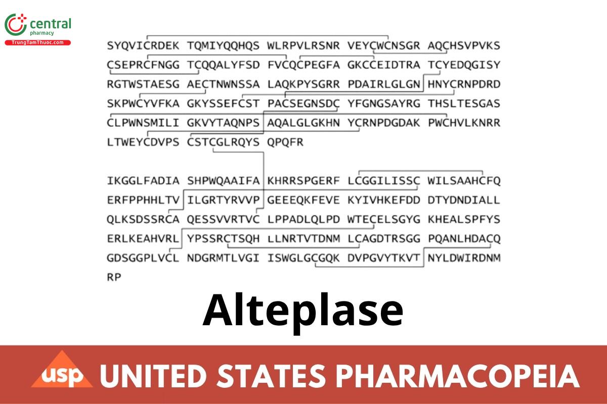
Alteplase is a highly purified glycosylated serine protease with fibrin-binding properties and plasminogen-specific proteolytic activities. It is produced by recombinant DNA synthesis in mammalian cell culture. It has a biological potency of NLT 90.0% and NMT 115.0% of the potency stated on the label, the potency being 580,000 USP Alteplase Units/mg of protein.
The presence of host cell DNA and host cell protein impurities in Alteplase is process specific; the limits of these impurities are determined by validated methods.
2 IDENTIFICATION
2.1 A
Standard solution: 1.0-2.5 mg/mL of USP Alteplase RS
Sample solution: Prepare similarly to the Standard solution.
Analysis
Samples: Standard solution and Sample solution
To each of three test tubes transfer 1 mL of 0.5-mg/mL H-D-isoleucyl-prolyl-arginyl-p-nitroaniline dihydrochloride. Separately transfer 200 µL of the Standard solution and 200 µL of the Sample solution to two of the test tubes. To the third test tube add 200 µL of 0.2 M arginine solution that has been adjusted with phosphoric acid to a pH of 7.3 (negative control). Mix the solutions in the three test tubes, and allow to stand for 1 min.
Acceptance criteria: A yellow color is produced in the solutions from the Standard solution and the Sample solution, while no yellow color is produced in the negative control.
2.2 B. PEPTIDE MAPPING
Solution A: 6.9 mg/mL of monobasic sodium phosphate in water, adjusted with phosphoric acid to a pH of 2.85. Filter, and degas.
Solution B: Acetonitrile
Mobile phase: See Table 1.
Table 1
| Time (min) | Solution A (%) | Solution B (%) |
| 0 | 100 | 0 |
| 91 | 70 | 30 |
| 121 | 40 | 60 |
| 131 | 40 | 60 |
Dialysis solution: 480 mg/mL of Urea, 44 mg/mL of tris(hydroxymethyl)aminomethane, and 0.88 mg/mL of edetic acid in water. Adjust with hydrochloric acid to a pH of 8.6.
Standard solution: Prepare a solution containing 1.0 mg/mL of USP Alteplase RS in water. Dialyze 2.0 mL of this solution into the Dialysis solution at room temperature for NLT 12 h. Measure the volume of the solution, and transfer it to a clean test tube. For each mL of solution in the tube, add 10 µL of 1 M dithiothreitol. Incubate at room temperature for 4 h, then add 25 µL of 1 M iodoacetic acid per mL of the solution, and incubate in the dark for 30 min. Quench the reaction by adding 50 µL of 1 M dithiothreitol per mL of the solution. Dialyze the solution against 0.1 M ammonium bicarbonate for 24 h, replacing the 0.1 M ammonium bicarbonate twice during the dialysis period. To 2.0 mL of the dialyzed solution, add 20 µg of trypsin, and incubate for 6-8 h at room temperature. Again add 20 µg of trypsin, and incubate for 16-18 h for a total of 24 h of incubation of the trypsin-treated solution. [NOTE-Store the Standard solution in a freezer.]
Sample solution: Using an accurately weighed quantity of Alteplase, proceed as directed in the Standard solution.
Chromatographic system
(See Chromatography (621), System Suitability.)
Mode: LC
Detector: UV 214 nm
Column: 4.6-mm x 10-cm; packing L1
Flow rate: 1 mL/min
Injection volume: 100 µL
System suitability
Sample: Standard solution
Suitability requirements
Resolution: NLT 1.5 between peaks 6 and 7 as defined by the USP Alteplase RS Data Sheet. The times for peaks 6 and 7 baseline widths are NMT 0.5 min.
Analysis
Samples: Standard solution, Sample solution, and a mixture of the Standard solution and the Sample solution (1:1) Measure the responses for NLT 20 major peaks as defined in the USP Alteplase RS Data Sheet.
Acceptance criteria: The retention times of the corresponding peaks of the Standard solution and the Sample solution do not differ by more than 0.4 min, and the peak area ratios relative to peak 19 (as shown on the USP Alteplase RS Data Sheet) do not differ by more than 20%. No additional significant peaks or shoulders are found, a significant peak or shoulder being defined as one having a peak response of NLT 5% of peak 19.
3 ASSAY
BIOLOGICAL POTENCY
Buffer: 1.38 mg/mL of monobasic sodium phosphate, 7.10 mg/mL of anhydrous dibasic sodium phosphate, 0.20 mg/mL of sodium azide, and 0.10 mg/mL of Polysorbate 80 in water
Human thrombin solution: 33 U.S. Units in terms of the U.S. Standard Thrombin/mL in Buffer
Human fibrinogen solution: 2 mg/mL of human fibrinogen in Buffer
Human plasminogen solution: 1 mg/mL of human plasminogen in Buffer
Standard stock solution: 1.0 mg/mL (580,000 USP Alteplase Units) of USP Alteplase RS in water
Standard solutions: Dilute volumes of Standard stock solution with water to obtain a series of five Standard solutions having known concentrations ranging from 145 to 9.3 USP Alteplase Units/mL.
Sample stock solution: 1.0 mg/mL of Alteplase in water
Sample solutions: Dilute a volume of Sample stock solution with Buffer to obtain a series of dilutions of about 1:20,000; 1:10,000; and 1:5,000.
Analysis
Samples: Standard solutions and Sample solutions
To a set of labeled glass test tubes add 0.5 mL of Human thrombin solution. To separate test tubes add 0.5 mL of each Standard solution or Sample solution, and store on ice. To a second set of labeled glass tubes, add 20 µL of Human plasminogen solution and 1 mL of Human fibrinogen solution, and store on ice. Beginning with the thrombin-Standard solution mixture containing the Standard solution with the lowest number of USP Units/mL, record the time, and separately add 200 µL of each of the thrombin-Standard solution mixtures to the test tubes containing the plasminogen-fibrinogen mixture. Using a vortex mixer, intermittently mix the contents of each tube for a total of 15 s, and carefully place into a rack in a 37° circulating water bath. A visually turbid clot forms within 30 s, followed by the formation of bubbles within the clot. Record the clot lysis time (tcl) from the first addition of the Alteplase solution to the last bubble to rise to the surface.
Using a least squares fit, determine the equation of the line using the log values of the standard concentration, in USP Alteplase Units/mL, versus the log values of their clot lysis times in s taken:
log t = m(logUS) + b
t = time to bubble release (s)
m = slope of the line
US = activity of the Standard solution (USP Alteplase Units/mL)
b = y-intercept of the line
The correlation coefficient is NLT -0.9900. From the line equation and using the log of the clot lysis time for the Sample solution, calculate the log of the activity (UA):
log UA = {[(log t) - b]/m}
Calculate the alteplase activity in USP Alteplase Units/mL taken:
Result = D(10logU)
D dilution factor for the Sample solution
Calculate the specific activity in the portion of Alteplase taken:
Result = (UA/P)
P = concentration of protein obtained in the test for Protein Content
Acceptance criteria: 90.0%-115.0% of the potency stated on the label, the potency being 580,000 USP Alteplase Units/mg of protein
4 OTHER COMPONENTS
PROTEIN CONTENT
Arginine solution: 34.8 mg/mL of arginine in water. Adjust with phosphoric acid to a pH of 7.3.
Sample stock solution: 1 mg/mL of Alteplase in water
Sample solution: Dilute a volume of Sample stock solution with a volume of Arginine solution to obtain a solution having an absorbance value of 0.5-1.0 at the wavelength of maximum absorbance at about 280 nm. Determine the dilution volume (V).
Instrumental conditions
(See Ultraviolet-Visible Spectroscopy (857).)
Mode: UV
Wavelength range: 240-500 nm
Analytical wavelengths: 320 nm and maximum absorbance at about 280 nm
Cell: 1 cm
Blank: Arginine solution
Analysis
Samples: Sample solution and Blank
Calculate the protein content in the portion of Alteplase taken:
Result = [(Amax - A320)/ε] x V
Amax = absorbance value at the wavelength of maximum absorbance
A320 = absorbance of the Sample solution at 320 nm
ε = molar absorptivity of Alteplase, 1.9
V = volume of Arginine solution required to prepare the Sample solution
5 IMPURITIES
CHROMATOGRAPHIC PURITY
SDS buffer: 400 mg/mL of glycerol, 5.52 mg/mL of tris(hydroxymethyl)aminomethane hydrochloride, 3.28 mg/mL of tris(hydroxymethyl)aminomethane, 0.20 mg/mL of bromophenol blue, and 0.20 mg/mL of xylene cyanole FF in sodium dodecyl sulfate solution (8 in 100)
Diluted SDS buffer: Dilute 1 volume of SDS buffer with 4 volumes of water.
Running buffer: 3.03 mg/mL of tris(hydroxymethyl)aminomethane and 14.26 mg/mL of glycine in sodium dodecyl sulfate (1 in 1000)
Carboxymethylation buffer: 480 mg/mL of urea, 44 mg/mL of tris(hydroxymethyl)aminomethane, and 1.2 mg/mL of edetic acid in water.
Adjust with hydrochloric acid, if necessary, to a pH of 8.6.
Ammoniacal silver nitrate solution: Transfer 105 mL of sodium hydroxide solution (0.36 in 100) and 7.0 mL of ammonium hydroxide to a 500-mL volumetric flask, and add slowly, with stirring, 20.0 mL of silver nitrate solution (20 in 100). Dilute with water to volume. [NOTE-Prepare this solution immediately before use, and protect it from light. This amount of solution is sufficient for two slab gels.]
Citric acid-formaldehyde solution: To 500 mL of water add 25 mg of citric acid, 0.25 mL of formaldehyde, and 0.025 mL of methanol, omitting the methanol if the formaldehyde is preserved with methanol. [NOTE-Prepare this solution fresh at the time of use. This amount of solution is sufficient for two slab gels.]
Gel: Prepare a 10% T (total acrylamide)-0.25% C (cross-linked bisacrylamide) resolving gel containing 0.1% sodium dodecyl sulfate, 0.375 M tris(hydroxymethyl)aminomethane hydrochloride, and 0.05 M tris(hydroxymethyl)aminomethane.
Arginine solution: 34.8 mg/mL of arginine in water. Adjust with phosphoric acid to a pH of 7.3.
Molecular weight standard solution: Use a commercially available preparation of low molecular weight protein standards (10,000-100,000 Da) at 2 mg/mL. Mix 990 µL of Diluted SDS buffer and 10 µL of the molecular weight standard mixture.
Control solution: Prepare a control solution of bovine serum Albumin containing 10 µg/mL. For a 10 ng/25 µL load, mix 600 µL of Diluted SDS buffer and 25 µL of the control solution, and heat at 90° for 2 min. For a 2.5 ng/25 µL load, mix 594 µL of Diluted SDS buffer and 6 µL of the control solution, and heat at 90° for 2 min.
Standard stock solution: 1 mg/mL of USP Alteplase RS in water
Standard solution: Dilute an accurately measured volume of Standard stock solution in Arginine solution to obtain a solution having a final concentration of 0.25 mg/mL. Heat 0.5 mL of this solution with 116 µL of SDS buffer and 10 µL of 1 M dithiothreitol at 80° for 2 min.
Carboxymethylated standard solution: Dilute 1.0 mL of Standard stock solution with 1 mL of Carboxymethylation buffer, and adjust with 1 M sodium hydroxide to a pH of 8.5. Add 20 µL of 1 M dithiothreitol, and incubate at 37° for 60 min. Add 100 µL of 1 M iodoacetic acid, and incubate in the dark for 20 min. Desalt by passing the solution through a chromatographic column containing fine gel chromatographic packing equilibrated with a buffer solution containing 20 mg/mL of sodium dodecyl sulfate, 100 mg/mL of glycerol, 1.42 mg/mL of tris(hydroxymethyl)aminomethane hydrochloride, and 0.85 mg/mL of tris(hydroxymeth yl)aminomethane. Collect the protein fraction of the preparation by elution with the same buffer, and add 20 µL of 1 M dithiothreitol. Adjust the protein concentration to about 0.2 mg/mL. with a buffer solution containing 20 mg/mL of sodium dodecyl sulfate, 100 mg/mL of glycerol, 1.42 mg/ml. of tris(hydroxymeth yl)aminomethane hydrochloride, 0.85 mg/mL of tris(hydroxymethyl)aminomethane, 1.06 mg/mL of dithiothreitol, 0.05 mg/mL of bromophenol blue, and 0.05 mg/mL of xylene cyanole FF.
Sample stock solution, Sample solution, and Carboxymethylated sample solution: Using an accurately weighed quantity of Alteplase, proceed as directed for Standard stock solution, Standard solution, and Carboxymethylated standard solution.
Blank: Mix 500 µL of water, 126 µL of SDS buffer, and 10 µL of 1 M dithiothreitol.
Analysis
Samples: Molecular weight standard solution, Control solution, Standard solution, Carboxymethylated standard solution, Sample solution, Carboxymethylated sample solution, and Blank
Separately apply equal volumes (about 25 µL) of the Standard solution, Carboxymethylated standard solution, Sample solution, and Carboxymethylated sample solution at the 5-µg load; apply equal volumes (about 38 µL) of the Standard solution and the Carboxymethylated standard solution at the 7.5-µg load; and apply the Control solutions at the 10-and 2.5-ng load onto separate lanes of the gel. Apply about 25 µL of the Molecular weight standard solution to each side of the gel, and apply about 25 µL of the Blank onto a separate lane. Apply the Standard solution and the Sample solution on one half and the Carboxymethylated standard solution and Carboxymethylated sample solution on the other half. Perform the electrophoresis using a constant current of 1.3-1.5 mAmp/cm of gel length and the Running buffer. Remove the gel from the apparatus 10-20 min after the tracking dye starts to move. Place the gel in 250 mL of a solution of 20% alcohol and 6% glacial acetic acid for NLT 1 h, and change the solution every 20 min, leaving the gel to soak overnight following the last change.
Perform silver staining of the gel by placing the gel in 250 mL of a 10% glutaraldehyde solution (v/v) in a shallow dish, and shake for about 30 min. Replace the glutaraldehyde solution with distilled water, allow the gel to soak for about 20 min, and then change the water. Repeat for a total of three washings. Transfer the gel to a dish, and cover with 250 mL of Ammoniacal silver nitrate solution. Place the dish on a shaker for about 15 min. Rinse four times with 250 mL of water, rocking the dish for 1 min between rinses. Continue rocking to prevent the gel from sticking and to facilitate washing.
Transfer the gel to a clear dish containing 250 mL of Citric acid-formaldehyde solution, and rock the dish. Protein bands become visible. When the gel is visibly stained, wash immediately with water, and rinse it repeatedly with water to remove the Citric acid-formaldehyde solution. Rinse the gel for NLT 1 h, and dry. Soak cellophane membranes in Glycerol solution (2 in 100). Roll a membrane onto a rigid sheet of plastic. Roll the gel onto the membrane, and cover with another membrane. Lay a frame on the edges of the membranes, and clamp it to the rigid plastic sheet. Dismantle the dryer, and cut off excess cellophane when dry (about 24 h). Visually examine the gel under light.
System suitability: The 2.5- and 10-ng Control solutions must be visible. The nonreduced Control solutions migrate with an apparent molecular weight of slightly less than 66,000 Da, as compared with the Molecular weight standard solution.
Acceptance criteria: The Sample solution exhibits three major bands in the region between 66,000 Da and 31,000 Da, corresponding to the major bands from the Standard solution. The Carboxymethylated sample solution exhibits six major bands in the region between 92,500 Da and 45,000 Da, corresponding to the major bands from the Carboxymethylated standard solution.
6 SPECIFIC TESTS
6.1 BACTERIAL ENDOTOXINS TEST (85)
NMT 1 USP Endotoxin Unit/mg of Alteplase
6.2 SINGLE-CHAIN CONTENT
Mobile phase: 27.6 g of monobasic sodium phosphate in 1000 mL of sodium dodecyl sulfate solution (1 in 1000). Adjust with sodium
hydroxide to a pH of 6.8. Filter, and degas.
Dithiothreitol solution: 3.12 mg/mL of dithiothreitol in Mobile phase
Standard stock solution: Using an accurately weighed quantity of USP Alteplase RS, make a 1-mg/mL solution in water.
Standard solution: Pipet 1 mL of the Standard stock solution into a glass tube, add 3 mL of Dithiothreitol solution, cap the tube, and invert to mix. Heat for 3-5 min at about 80°.
Sample stock solution: Using an accurately weighed quantity of Alteplase, make a 1-mg/mL solution in water.
Sample solution: Pipet 1 mL of the Sample stock solution into a glass tube, add 3 mL of Dithiothreitol solution, cap the tube, and invert to mix. Heat for 3-5 min at about 80°.
Chromatographic system
(See Chromatography (621), System Suitability.)
Mode: LC
Detector: UV 214 nm
Column: 7.5-mm x 60-cm; packing L25
Flow rate: 0.5 mL/min
Injection volume: 50 µL
System suitability
Sample: Standard solution
Suitability requirements
Resolution: NLT 1.1 between the single-chain and two-chain alteplase peaks
Analysis
Samples: Standard solution and Sample solution
[NOTE-The major peaks are from single-chain and two-chain alteplase and from higher and lower molecular weight species.]
Calculate the percentage of single-chain alteplase in the portion of Alteplase taken:
Result = (rU/rT) × 100
rU = peak response for single-chain alteplase
rT = sum of all the peak responses of alteplase
Acceptance criteria: No peaks or shoulders in the Sample solution that are not present in the Standard solution are found; NLT 60%.
7 ADDITIONAL REQUIREMENTS
PACKAGING AND STORAGE: Preserve in tight containers, and store in the frozen state at a temperature of -20° or below.
USP REFERENCE STANDARDS (11)
USP Alteplase RS
